News — 24/04/2018
Optos – The difference in Retinal Imaging
Optos was founded by Douglas Anderson after his then five-year-old son Leif went blind in one eye when a retinal detachment was detected too late. The intention was to create a way of non-invasively capturing as much of the retinain one image as possible.
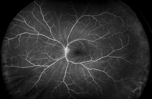
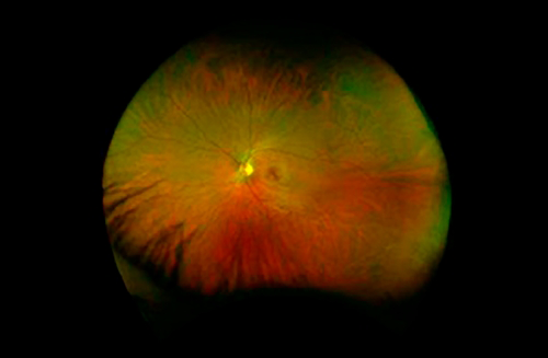
Regular eye exams are vital to maintaining your vision and overall health. At Eyes of Claremont we offer the optomap® as an important part of our eye exams. The optomap produces an image that is unique and provides us with a high-resolution 200° image to check the health of your retina. This is much wider than a traditional 45° image. Many eye problems can develop without you knowing – in fact, you may not even notice any change in your sight. Fortunately, diseases or damage such as macular degeneration, glaucoma, retinal tears or detachments, and other health problems such as diabetes and high blood pressure, can be seen with a thorough exam of the retina.
The inclusion of optomap as part of a comprehensive eye exam provides:
- A scan to show a healthy eye or detect disease.
- A view of the retina, giving us a more comprehensive view than other methods.
- The opportunity for you to view and discuss the optomap image of your eye with us at the time of your exam.
- A permanent record for your file, which allows us to view your images over time and look for changes.
The optomap is fast, easy, and comfortable for anyone. The entire image process consists of you looking into the device one eye at a time. The optomap images are shown immediately on a computer screen so we can review it with you.


